


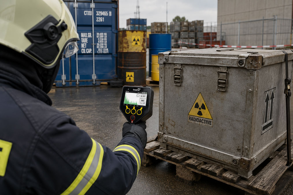
A radionuclide identifier initially reports Cs-137. After a longer measurement, a second nuclide appears with lower confidence. At another measurement position, the spectrum changes again. Are several radioactive sources actually present, or is the geometry causing an incorrect automatic identification?
Such situations are among the more demanding tasks in mobile gamma spectrometry. A radionuclide identifier, or RID, not only evaluates the dose rate but also analyses the measured energy spectrum and compares characteristic structures with a stored nuclide library.
With a single unshielded source and sufficient counting statistics, this task is comparatively straightforward. It becomes more difficult with several radionuclides, shielding material, a high natural background or unfavourable measurement geometry. Characteristic lines can then overlap, be attenuated to different degrees or be partially obscured by scattered radiation.
Radionuclide identifiers for corresponding applications can be found under Radionuclide Identifiers. Further instruments for dose rate measurement, contamination monitoring and radiation protection are grouped under Radiation Measurement Technology.
Table of Contents
- How does an RID identify a radionuclide?
- What happens when several radionuclides are present?
- How does shielding alter the gamma spectrum?
- What role does energy resolution play?
- Correctly assessing background and NORM
- Choosing the correct distance and measurement geometry
- Why does a longer measurement time only help to a certain extent?
- Assessing automatic identification and confidence
- Dose rate takes priority over spectrum optimisation
- Systematically rechecking an uncertain identification
- Practical example of a shielded mixed source
- Which RID is suitable?
- Conclusion
- Frequently asked questions
How does an RID identify a radionuclide?
Many radionuclides emit gamma photons with characteristic energies. A spectrometric detector therefore does not simply produce a total count rate; it also assigns the detected events to their respective energies. This produces a gamma spectrum.
In an idealised case, characteristic gamma energies appear as peaks. The RID’s internal software searches for these structures and compares their energies, intensities and, in some cases, the shape of the entire spectrum with known radionuclides stored in a library.
Identification is therefore not based on a single indication. A capable algorithm must simultaneously assess which lines are present, which should theoretically be present and whether the overall spectrum statistically corresponds to one or more radionuclides.
This is precisely why mixed and shielded sources are challenging: the actual measured spectrum can look significantly different from an ideal reference spectrum.
What happens when several radionuclides are present?
If two or more radionuclides are simultaneously within the detector’s field of view, their spectra are physically superimposed. The RID does not see two separate measurements but one combined spectrum.
Gamma energies that are clearly separated from one another can often still be assigned reliably. The situation becomes more difficult when lines from different nuclides are close together or when a strong source spectrally dominates a weaker source.
Another problem results from different activities and distances. A weak source located directly next to the detector can contribute more strongly than a significantly more active source that is farther away or more heavily shielded.
The automatic nuclide indication should therefore not be regarded as a simple list of all substances actually present. It is the result of the spectrum that the detector was able to record under the current measurement geometry.
How does shielding alter the gamma spectrum?
Shielding frequently leads to misinterpretations. An important distinction must be made here: the characteristic energy of a directly detected gamma photon is not simply shifted by shielding located in front of the source.
Instead, photons are attenuated to different degrees depending on their energy, the shielding material and the material thickness. Lower energies in particular can be suppressed much more strongly. In addition, scattering processes generate photons with lower energies, increasing the background or Compton continuum.
The spectrum behind shielding can therefore exhibit completely different relative intensities compared with the spectrum of the same source without shielding.
| Influence | Effect on the Spectrum | Possible Consequence for RID Evaluation |
|---|---|---|
| Metal shielding | Certain energy ranges are attenuated more strongly | Important identification lines can become very weak |
| Compton scattering | Increased continuous background below the original gamma energy | Small peaks become more difficult to detect |
| Several radionuclides | Spectra overlap | Lines can be assigned to several nuclides |
| Strongly differing source strengths | One source dominates the spectrum | Weaker nuclides may only become visible after a longer measurement |
| Change in geometry | Relative source contributions change | Identification may change between measurement positions |
Real-world applications show that this is not merely a theoretical issue. With heavily shielded industrial sources, the measured spectrum can at times be dominated mainly by scattered radiation, making reliable automatic identification considerably more difficult.
What role does energy resolution play?
A detector does not represent a specific gamma energy as an infinitely narrow line. Every peak has a measurable width. How well two neighbouring lines can be separated depends to a significant extent on the detector’s energy resolution.
High detection efficiency and high energy resolution are different characteristics. A large scintillation detector can register many photons and therefore achieve good counting statistics quickly. A high-resolution laboratory spectrometer, by contrast, can separate closely spaced lines more effectively.
A mobile RID therefore always represents a compromise between sensitivity, robustness, size, measurement speed and spectral resolution.
This also means that a longer measurement time can improve a low number of detected events, but it cannot arbitrarily separate two peaks that cannot be physically resolved sufficiently by the detector.
Correctly assessing background and NORM
An RID never measures only the suspicious source. The natural and technical radiation environment is always detected at the same time.
Naturally occurring radioactive materials, commonly referred to as NORM, can for example generate contributions from natural uranium and thorium decay series or potassium-40. Depending on the building material, soil, mineral or cargo, this contribution can be significantly above the usual local background.
Medical or industrial radionuclides may also be present. An alarm in the vicinity of a hospital, airport or logistics centre must therefore be assessed in a different context from the same measured value at a remote storage area.
A good background measurement helps the RID make additional spectral structures more visible. Especially with weak sources, it is therefore important to know whether the observed lines actually belong to the source being investigated or were already present beforehand.
However, in the case of an unknown find, the background should only be measured from an appropriate safe position. The suspicious source should not be moved or opened solely to improve the spectrum.
Choosing the correct distance and measurement geometry
Distance has a major influence on the measured count rate. At the same time, with mixed sources, even a change in measurement position can alter the relative contributions of individual radionuclides.
This can be diagnostically useful. If, for example, significantly different spectra are observed from two safe measurement positions, this may indicate that several spatially separated sources are present within the measurement area.
However, the smallest possible distance is not automatically the correct choice. With a strong source, an excessively high count rate can burden the detector or spectral evaluation. More importantly, radiation protection requirements always take priority over obtaining the best possible statistical quality.
The measurement distance should therefore be selected according to the dose rate, the properties of the instrument and the intended operating procedure.
Why does a longer measurement time only help to a certain extent?
With weak radiation, only a very small number of events may be present in a characteristic peak after a few seconds. A longer measurement time then improves statistical reliability.
As the number of recorded events increases, the signal of a genuinely present line becomes easier to distinguish from the random statistical background.
A longer measurement can therefore turn an initially uncertain identification into a significantly more stable assessment.
However, it does not solve every problem. If a characteristic low-energy line is almost completely absorbed by heavy shielding, even a considerably longer measurement may only help to a limited extent. The same applies if two peaks can barely be separated because of the detector’s energy resolution.
Measurement time should therefore not be optimised in isolation. Geometry, background, count rate and spectral shape must be considered together.
Assessing automatic identification and confidence
An automatic nuclide indication is a decision made by the evaluation algorithm on the basis of the available data. With a clean spectrum, the result can be very stable. With mixed sources, poor statistics or heavy shielding, uncertainty increases.
For this reason, the displayed nuclide name should not be considered in isolation. Where the instrument provides such information, confidence, spectrum, measurement duration, count rate and any additional nuclide candidates are also relevant.
Particular attention should be paid when the identification changes repeatedly between several directly consecutive measurements. This can indicate insufficient statistics, changing measurement geometry, a mixed source or a heavily disturbed spectrum.
Low confidence does not automatically mean that the suggested nuclide is incorrect. Rather, it means that the measured data does not support the assignment as clearly as it would for an unambiguous single source.
Dose rate takes priority over spectrum optimisation
With an unknown radioactive source, perfect radionuclide identification is not the first priority. The dose rate and the radiation protection situation must be assessed first.
A measurement position should not be moved closer to the source solely because this would produce a better spectrum. Likewise, unknown shielding should not be opened or removed merely to make hidden gamma lines visible.
The basic principles of minimising exposure time, maximising distance and using shielding remain paramount. Work involving unknown or potentially highly active sources should only be carried out by appropriately trained and authorised personnel.
An RID is an important decision-support instrument in this context. However, it does not replace the organisation’s radiation protection procedures and is not automatically an official personal dosimeter.
Systematically rechecking an uncertain identification
When the nuclide indication is contradictory, a structured follow-up measurement is more useful than repeatedly trying random measurement positions.
| Test Step | Observation | Assessment |
|---|---|---|
| Check dose rate | Is the current measurement position acceptable from a radiation protection perspective? | Safety determines the permissible measurement geometry |
| Measure background | Which lines are already present without the suspicious source? | Distinguish natural or local background |
| Repeat measurement | Does the nuclide indication remain stable? | Identify an individual statistical outlier |
| Extend measurement time | Do weak peaks become more distinct? | Assess insufficient counting statistics |
| Use a safe second position | Do the relative peak intensities change? | Indication of geometry, shielding or several sources |
| Save spectrum and identification list | Which candidates and spectral data are available? | Enable subsequent expert evaluation |
| Escalate if uncertainty remains | Identification remains unclear or is safety-relevant | Arrange high-resolution or specialist follow-up measurement |
Automatic RID indications should not be overinterpreted, particularly when safety-related decisions are involved. If the spectrum remains ambiguous, subsequent high-resolution gamma spectrometry may be required.
Practical example of a shielded mixed source
During an inspection, an elevated gamma dose rate is detected at a metal container. After a short measurement, the RID indicates an industrial radionuclide, but the confidence fluctuates. A second nuclide candidate also appears intermittently.
The first response should not be to open the container or remove any shielding that is present.
Instead, the dose rate is first documented from a safe position. A longer spectrometric measurement is then carried out. This makes the dominant peak statistically much more stable, while a second weaker structure also becomes visible.
At another permissible measurement position, the ratio between the two spectral contributions changes. This indicates that the original spectrum may not simply originate from one homogeneous single source.
At the same time, the spectral background shows a pronounced scattering component. Existing metal shielding therefore significantly alters the observed intensities.
The result is therefore not interpreted as a final and unambiguous identification. The spectrum, dose rate, measurement position and automatic nuclide candidates are stored and made available for further expert assessment.
This example demonstrates the key point: an RID provides valuable information under difficult measurement conditions, but the quality of the decision depends on evaluating spectrum, geometry and confidence together.
Which RID is suitable?
GRAETZ RadXplore-ident
The GRAETZ RadXplore-ident is suitable, for example, for mobile applications involving fire brigades, customs authorities, radiation protection personnel and security-related inspections.
The instrument combines a 2 × 1 inch BGO detector with high-speed digital electronics. The relatively large detector provides high detection efficiency, which is particularly advantageous for rapid on-site measurements and weaker sources.
The RadXplore-ident has a nuclide library containing more than 70 radionuclides and can distinguish or identify natural, medical and industrial radiation sources. Dose rate measurement, source localisation and neutron detection are also available.
Spectra and measurement data can be stored for documentation purposes or further processed via digital interfaces. This is particularly important for complex mixed spectra because subsequent expert assessment can go beyond the simple indication shown on the display.
Under Radionuclide Identifiers, you will find corresponding instruments for detection, localisation and nuclide identification.
Further instruments for dose rate measurement, personal protection and contamination monitoring are grouped under Radiation Measurement Technology.
ICS Schneider Messtechnik provides support in selecting suitable radiation measuring instruments for fire brigades, industry, authorities, security applications and operational radiation protection.
Conclusion
Radionuclide identification becomes significantly more demanding when several sources, shielding or a high background are present at the same time.
With mixed sources, the gamma spectra overlap. A strong radionuclide can partially obscure the lines of a weaker source, while closely spaced gamma energies can only be separated to a limited extent depending on the detector’s energy resolution.
Shielding must also be interpreted correctly. It does not normally simply shift the characteristic gamma energies. Instead, it alters the relative intensities, attenuates certain energy ranges in particular and increases the spectral background through scattering.
A longer measurement time improves statistics and can make weak peaks visible. However, it cannot compensate for a completely suppressed line or a fundamentally insufficient separation of two closely spaced peaks.
An RID indication should therefore never be reduced to the name of the automatically detected nuclide alone. Measurement time, count rate, background, geometry, spectrum and confidence must all be considered together.
Radiation protection also takes priority when dealing with unknown sources. Shielding should not be removed without authorisation and measurement distances should not be reduced solely to improve the spectrum.
A modern RID is therefore primarily a powerful decision-support tool. In difficult mixed-source or shielding situations, additional high-resolution specialist spectral analysis may still be required.
Frequently asked questions about radionuclide identification in mixed radiation fields
Can an RID detect several radionuclides at the same time?
Yes. Suitable evaluation algorithms can analyse mixed spectra and determine several nuclide candidates. However, reliability depends on factors including activity, measurement time, energy resolution, shielding and spectral overlap.
Does lead shift the gamma peaks of a radionuclide?
The original characteristic gamma energies are not simply shifted by the shielding. However, attenuation and scattering alter the observed spectrum, so peak intensities can change significantly or individual lines may become barely visible.
Why does the RID identify a different nuclide behind shielding?
Certain lines can be attenuated more strongly than others, while additional scattered radiation can increase the background. As a result, different spectral information is available to the identification algorithm.
Does a longer measurement time always help?
A longer measurement improves counting statistics and is particularly helpful with weak sources. However, it cannot fully compensate for insufficient energy resolution or heavily absorbed gamma lines.
What does a fluctuating nuclide identification mean?
It can indicate poor statistics, a mixed spectrum, changing geometry, background influences or shielding. In such cases, the spectrum and measurement conditions should be considered in addition to the automatic indication.
What is NORM?
NORM stands for naturally occurring radioactive material. Elevated concentrations of natural radionuclides can occur, for example, in minerals, ores, building materials or industrial residues and can influence an RID measurement.
Should shielding be removed to improve identification?
Not in the case of an unknown source without appropriate authorisation and radiation protection assessment. Improving the spectrum must not result in increased exposure or unsafe handling of the source.
When is a high-resolution follow-up measurement advisable?
If several radionuclides cannot be clearly separated, automatic identification remains unstable or a safety-related decision depends on the result, further high-resolution gamma spectrometry may be appropriate.